Suite 206 4938 Brownsboro Road Louisville, KY 40222


M-F: 8:00am – 4:30pm (502) 339-2922  

Moles affects about
327 million
people in the U.S.
People with 10 or more
atypical moles
have 12X the risk of Melanoma
A common mole is usually
smaller than
5mm wide


Having +50 common moles
increases
the risk of Melanoma

What are Moles?

Moles, also called nevi, are an extremely common type of skin growth. They can develop anywhere on the body and are typically brown in color, although they can also appear black, tan, pink, blue, or red. Most moles have an oval or round shape, and they commonly develop during childhood or adolescence.

It’s normal for some moles to have hair growing from them, and it’s common for some moles to lighten or darken in color or even fade away over time. Although most moles are harmless, you should always monitor them for any changes in their appearance. This is an important step in the early detection of cancerous moles. Melanoma is the most serious form of skin cancer.

Are There Different Types of Moles?

There are three types of moles that are the most common. Let’s take a closer look at each type:

Congenital mole: A congenital mole is any type of mole you’re born with. They can vary in color and are typically flat in appearance. According to the American Academy of Dermatology (AAD), having a congenital mole increases your risk of developing melanoma.

Atypical mole: This type of mole is also called a dysplastic nevus and is larger in size than a common mole. They often have irregular-shaped borders and can be more than one color. These moles can appear on most areas of the body, including your torso, scalp, and neck.

Common mole: Common moles, known as acquired moles, develop on the skin after birth. Most people typically have around 10-40 common moles, and they’re usually found on areas of the skin that are commonly exposed to sunlight.

How Do I Know If a Mole Is Cancerous?

Although most moles are harmless, melanoma can occur in a mole. This is why it’s important to know your skin and become familiar with the appearance of any moles you have. If you notice any changes in a mole, it’s important to bring it to the attention of your primary care provider or dermatologist. Melanoma is very treatable when caught early.

According to the American Cancer Society, an atypical mole is more likely to become cancerous in comparison to a common mole. Cancerous moles can vary in appearance. Your doctor should evaluate moles that feel painful, tender, itch, bleed, or ooze. You should also have moles checked if you notice any difference in a mole’s shape, color, size, or height. The AAD also recommends visiting a dermatologist for an evaluation if you develop a new mole anywhere on your body after age 30.

Moles develop when cells in the skin grow in large clumps or clusters instead of spreading evenly throughout the skin. These skin cells are called melanocytes, and they’re responsible for producing melanin, which gives your skin its pigment. Anyone can develop moles, but these factors can increase the likelihood of developing them:

  • Genetics
  • Age
  • Hormonal changes, such as pregnancy
  • Frequent sun exposure

When Is Mole Removal Necessary?

Unless they’re cancerous, most moles don’t require treatment. You might choose to have a mole removed if it’s easily irritated or frequently snags on clothing or jewelry. Some people also choose to have a mole removed for cosmetic reasons, such as a large or noticeable mole on the face or neck.

Remember, a dermatologist or other healthcare professional should always perform mole removal. Never attempt to remove a mole on your own. If you’re asking friends or family, “Is there a trusted doctor for mole removal near me?”, look no further than our office. Your doctor may recommend one of these treatment options:

Surgical shave: Using a surgical blade, the dermatologist will remove the mole at the surface.

Surgical excision: During this procedure, the dermatologist will cut out the entire mole. If needed, the area will be closed with stitches.

If a mole is removed with a surgical shave or a surgical excision, a doctor may examine the mole closely under a microscope to detect cancerous cells.

How Much Does Mole Removal Cost?

The mole removal cost can vary greatly and depends on a variety of factors. Insurance usually covers mole removal for medical reasons, including those that are precancerous or cancerous. Most moles removed for cosmetic reasons typically aren’t covered by insurance. The total cost for removal is also influenced by the mole’s size and location on the body.

Where Can I Find a Doctor Who Performs Mole Removal Near Me?

If you’re wondering, “What kind of doctor offers mole removal near me?”, it’s important that you seek care from a board-certified dermatologist. These specialists receive extensive training that enables them to safely remove moles and minimize scarring. They can also carefully examine moles for the early signs of melanoma.

This year approximately
100,350 new
melanoma cases will be diagnosed
Skin cancer is the
most common
among all cancers
You have higher risk if a
close relative
had melanoma
Using tanning beds increases
the risk
of melanoma by 75%

Is Melanoma Cancer?

Melanoma is the most dangerous and deadliest form of skin cancer. It begins in cells called melanocytes, which produce the pigment that gives skin its color. In comparison with other types of skin cancer, melanoma occurs less commonly, but it’s considered more serious. It can quickly spread throughout the body if melanoma treatment isn’t started early.

Your doctor or dermatologist should evaluate any change in an existing mole. You also need to frequently examine your skin for these melanoma symptoms:

  • An asymmetrical mole
  • A mole with an irregular border
  • A mole that changes color
  • A mole that is larger than a pencil eraser

Are There Different Types of Melanoma?

Melanoma cancer has four different types. Here’s what you need to know about each:

Lentigo maligna: This type of melanoma usually affects older people who have sun-damaged skin. It commonly develops on the face, ears, upper torso, and arms. Lentigo maligna presents as a blotchy patch that has uneven borders. The patch can be flat or slightly raised with colors that are usually blue, black, tan, or dark brown.

Acral lentiginous melanoma: Acral lentiginous melanoma is the most common type of melanoma that affects African-Americans and Asians. It typically appears as a brown or black discoloration on the palms of the hands or bottom of the feet. It can also be found under the fingernails and toenails.

Nodular melanoma: Nodular melanoma is one of the most aggressive types of skin cancer. It grows rapidly and affects deeper layers of skin. For this reason, it’s often diagnosed at a later stage. The main sign of nodular melanoma is a bump that feels firm to the touch and is raised above the surface of the skin. The bump usually appears as black or blue, but it can also be red or pink in color.

Superficial spreading melanoma: This is the most common type of melanoma. One of the first melanoma symptoms associated with superficial spreading melanoma is an asymmetrical spot with irregular borders that appears flat or slightly raised. Color variations can range from black, white, brown, tan, pink, red, or blue. While it can form on most areas of the body, superficial spreading melanoma commonly appears on the upper back of men and women, on the legs in women, and on the torso in men.

Melanoma Causes and Risk Factors

Melanoma develops as a result of damaged DNA in melanocytes. This damage leads to the uncontrolled growth of skin cells. One of the primary causes of damage to the DNA in skin cells is excessive exposure to the sun’s ultraviolet light. Over time, this damage can cause mutations that may lead to skin cancer.

In addition to frequent sun exposure, there are other melanoma causes and risk factors associated with its development. These include:

  • Experiencing frequent sunburns
  • Family history of melanoma
  • Fair skin, including people who have light eyes and light- or red-colored hair
  • Frequently using tanning beds
  • Living at a high elevation or close to the equator
Melanoma Causes

Melanoma Stages

After your doctor or dermatologist makes a diagnosis of melanoma, the next step is to determine its stage and severity level. Melanoma stages range from 
0 to IV. Stages 0 and I are the most easily treated because the melanoma hasn’t penetrated into the deeper layers of skin.

Stage II melanoma is considered intermediate, and the tumors are usually deeper than 1 mm. Stage III melanoma is an advanced stage in which the tumor has spread to the lymph nodes. Once melanoma reaches stage IV, it has spread to other parts of the body, most often the GI tract, brain, lungs, liver, and bone.

Melanoma Treatment Options

Melanoma Treatment Options

There are many options available, but the melanoma treatment you receive depends on the type of melanoma you have and the stage of the disease. Surgery to remove the melanoma is the most common treatment for early stages of the disease. Mohs surgery, in which skin cancer is removed layer by layer, may also be a treatment option for some patients with melanoma.

Surgery is also an option for advanced stages of the disease. Deeper excisions of the tumor or removal of the lymph nodes may be needed for advanced melanoma. If melanoma has spread to the lymph nodes or other parts of the body, immunotherapy can be a treatment option. Immunotherapy triggers the immune system’s ability to attack cancer cells.

Chemotherapy is another treatment option used to destroy melanoma cancer cells. Radiation therapy kills cancer cells with high-energy X-ray beams, and it’s also used to reduce the chance of recurrence after melanoma has been removed. With targeted therapy, drugs and other medications target the specific weaknesses found in cancer cells. This is a newer form of treatment that has less severe side effects in compariso

What are Hives?

Hives, also known as urticaria, is a common dermatological condition characterized by raised, itchy, and often red welts or wheals on the skin. These welts can vary in size and shape and may appear anywhere on the body. Hives are usually caused by an allergic reaction, but they can also result from other triggers such as stress, heat, certain medications, infections, insect stings, or food allergies. The duration of an outbreak of hives may be as short as a few hours.

Hives develop when histamines are produced in the body as a reaction. Histamine production causes the tiny blood vessels, known as capillaries, to leak fluid. When the fluid accumulates under the skin, it causes the red bumps that are hives. Hives often itch uncomfortably.

There are several different types of hives, the most common of which include:

Caused by ingesting certain foods or medication or through infections. Insect bites may also be a cause.

  • Chronic urticaria and angioedema: Usually affects internal organs and the exact causes are unknown, except that it is allergy related.
  • Physical urticaria: Caused by direct physical stimulation of the skin, such as extreme heat or sun exposure, this form of hives develops in about an hour.
  • Dermatographism: Hives formed after aggressive stroking or scratching of the skin.

What are the symptoms of Hives?

The skin swellings associated with hives are called wheals and are usually round, pink or red bumps. The skin surrounding the wheals may also be red.

Symptoms of chronic urticaria and angioedema include muscle soreness, shortness of breath, vomiting, and diarrhea.

There are several different types of hives, the most common of which include:

  • Caused by ingesting certain foods or medication or through infections. Insect bites may also be a cause.
  • Chronic urticaria and angioedema: Usually affects internal organs and the exact causes are unknown, except that it is allergy related.
  • Physical urticaria: Caused by direct physical stimulation of the skin, such as extreme heat or sun exposure, this form of hives develops in about an hour.
  • Dermatographism: Hives formed after aggressive stroking or scratching of the skin.
Hives Itchy Skin

Who gets Hives?

Though allergic symptoms differ with each person, anyone who has allergic tendencies can get hives. There are no tests for hives, only for the allergens that may trigger them.

Hives Treatment Options

The best treatment for hives is to identify the allergic component and eliminate it from the patient’s lifestyle. In the case of medications, however, this may not be feasible. Antihistamines may provide temporary relief and seem to be preventive when taken regularly, not just when an outbreak occurs. For chronic hives, oral corticosteroids may be prescribed.

Eczema affects over
31 million
people in the U.S.
There are
7 types
of Eczema
Studies show
1 in 3 infants
will have Eczema
It is more common in 
females
than in males

What Is Eczema?

Eczema is a common skin condition that affects more than 31 million people in the United States, according to the National Eczema Association (NEA). It causes dry, scaly skin with redness and itching, and blisters or tiny bumps with a rash-like appearance. In severe cases, the skin may form painful cracks that bleed and form a crust. It’s very common for babies and young children to develop eczema on their cheeks and chin, but it can appear anywhere on the body and affect people of all ages. The NEA estimates that one in ten people will develop eczema at some point in their lives.

If you have eczema, living with the condition can be challenging and frustrating. It can greatly affect your quality of life, often leading to difficulty sleeping, low self-esteem, embarrassment, and avoiding social situations. People with eczema commonly experience flare-ups and periods of remission. During a flare, skin is actively itchy and very inflamed. During a period of remission, skin heals and there may not be any noticeable symptoms. Remission can last weeks, months, or even years. Eczema isn’t contagious and can’t be transmitted from person-to-person.

An Everyday Guide to Eczema

Eczema Symptoms

The symptoms of eczema won’t be the same for everyone. For most people with eczema, their skin is extremely itchy. Because the skin itches so much, people often scratch their skin until it bleeds. In addition to intensely itchy skin, these are the most common eczema symptoms to watch out for:

Skin redness

Dry, sensitive skin

Scaly rash

Swollen, inflamed skin

Bumps or blisters that bleed or ooze when scratched

Dark-colored patches of skin

Patches of skin that appear rough or leathery

Are There Different Types of Eczema?


According to the NEA, there are seven different types of eczema:

Atopic dermatitis: This is the most common type of eczema. It’s a chronic and inflammatory type of eczema that typically affects the backs of the knees and elbows and produces symptoms that are typically seen in most people with eczema, such as dry, scaly skin, itching, and redness. Atopic dermatitis usually affects people who have hay fever and asthma.

Contact dermatitis: Irritant or allergic contact dermatitis occurs when your skin comes into contact with allergens or an irritating substance, such as some soaps and fragrances, detergents, pollen or animal dander, wool, or nickel in jewelry. Contact dermatitis makes the skin inflamed, causing your skin to burn and itch.

Nummular eczema: Nummular eczema produces scaly, coin-shaped patches that are very itchy. It usually occurs on the legs and is thought to be triggered by insect bites and dry skin during the winter.

Neurodermatitis: Neurodermatitis is very similar in appearance to atopic dermatitis, and it affects about 12% of people in the US, according to the NEA. Symptoms of neurodermatitis include thick, scaly patches that form on the scalp, neck, shoulders, hands, ankles, and feet. These patches are very itchy and bleed easily when scratched.

Seborrheic dermatitis: This type of eczema usually forms on parts of the body that have a lot of sebaceous, or oil-producing, glands, such as the scalp, back, or eyebrows. When it affects the scalp, seborrheic dermatitis results in dandruff. In infants, it’s known as “cradle cap.”

Dyshidrotic eczema: This skin condition produces small, fluid-filled blisters on the soles of the feet, toes, palms, and fingers. It’s much more common in women and is usually caused by moist hands and feet, allergies, stress, or exposure to nickel (found in some jewelry), chromium salts (found in some paints, leather, and cement), and cobalt (found in metal-plated objects).

Stasis dermatitis: Stasis dermatitis usually affects the lower legs and typically affects the elderly. It’s caused by a circulation problem that affects blood flow, causing fluid to leak out of weakened veins and into the skin. Symptoms include swollen ankles, open red sores, pain, and itching.

What Causes Eczema?

A specific eczema cause hasn’t been found, although there are several theories. Eczema does have a genetic basis and tends to run in families. It’s also thought that people with eczema have an overactive immune system that causes an inflammatory response when triggered by a specific substance. This is what leads to red, itchy skin and the other symptoms associated with eczema.

Research has also shown that some people with eczema lack a protein in their skin called filaggrin, which is responsible for building a healthy protective barrier on the top layer of skin. Without enough filaggrin, skin dries out rapidly, making it difficult to retain proper moisture levels. This is one of the reasons why people with eczema have very dry, itchy skin.

While the exact eczema cause is unknown, researchers have found that exposure to certain substances or even situations can trigger a skin reaction, resulting in noticeable symptoms. Take a look at some of the most common eczema triggers and irritants that can lead to eczema symptoms:

Environmental Factors

  • Dry air
  • Pollen
  • Pet dander
  • Dust mites
  • Mold
  • Tobacco
  • Pollution
  • Sweating
  • Extremes in temperature, such as very hot or cold

Lifestyle Factors

  • Food allergies
  • Stress
  • Hormonal changes

Irritant Products

  • Fragrances and perfumes
  • Laundry detergents
  • Soaps
  • Cosmetics
  • Chlorine
  • Cleaning products and disinfectants
  • Wearing clothing made of wool or synthetic fabrics
  • Certain metals, such as nickel

Can Eczema be treated?

There isn’t an eczema cure, but there are treatments to help manage symptoms. No treatment option will work for everyone, and treatments can range from eczema medicine, such as prescription topical treatments, over-the-counter products, and lifestyle modifications. Eczema treatment must be consistent regardless of which type of eczema you have.

Let’s take a closer look at the different ways to treat eczema:

Skin care: Ensuring that your skin stays moist is important if you have eczema. Itching increases when your skin is dry, so using a moisturizer, cream, or ointment can help keep your skin moisturized. Applying these products several times a day, especially after showering or bathing while your skin is still slightly wet, can help to restore the moisture barrier and hydrate your skin. Look for products containing hyaluronic acid, ceramides, and coconut oil. Products labeled “for sensitive skin,” “hypoallergenic,” and “fragrance free” are key words to look for.

Avoid triggers: For many people with eczema, exposure to certain ingredients or substances can cause symptoms to flare up. Avoiding products containing alcohol, perfumes, dyes, or fragrance can be beneficial and keep the itching and redness under control. You can also avoid wearing wool and other irritating fabrics, choosing loose cotton clothing instead.

Treat the itch: Preventing and treating itching is an important component of eczema treatment. Using over-the-counter creams with hydrocortisone as an active ingredient can help control the itching, swelling, and redness that occurs in people with eczema. For severe cases, stronger eczema medicine containing steroids can be prescribed by your dermatologist.

Although an eczema cure isn’t available, it’s possible to effectively control and minimize your symptoms with proper treatment and by practicing good skin care. You can also prevent flare-ups by resisting scratching, avoiding hot showers and baths, and minimizing stress.

Cold Sores – the virus that never leaves

Once infected with the HSV-1 cold sore virus, the virus never leaves your body. Although most people aren’t exactly sure when they first encounter the virus, it’s usually contracted in early childhood, even if it did not manifest as a visible cold sore. The virus will remain dormant until it is triggered. Cold sore triggers may include colds, flu, fever, stress, fatigue, and environmental factors like sun and wind.

What are the signs of a cold sore?

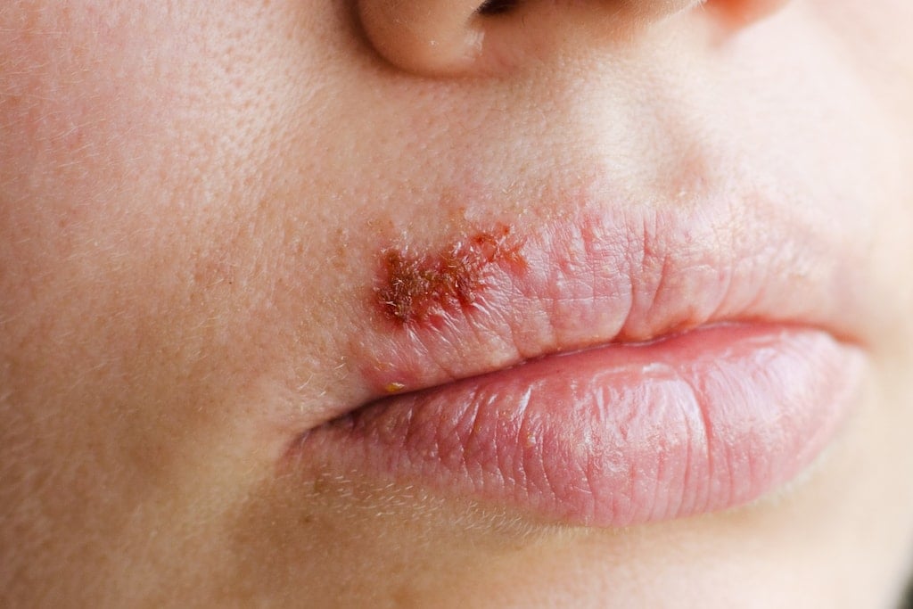
A cold sore can start as a burning or tingling sensation on your lip or around your mouth. It can develop into a small red patch with a blister. Once the blister opens, a hard crust will form and eventually fall off. The area affected will return to normal once the life cycle of a cold sore is complete. The life cycle of a cold sore is typically 10 to 14 days.

Are cold sores contagious?

Yes, cold sores are highly contagious. You can become infected with the virus by kissing, touching your fingers to your mouth or eyes (if your fingers are already infected), and sharing infected items. Sanitizing surfaces and good hand-washing practices can help eliminate the likelihood of spread.

How do you treat a cold sore?

Cold sores cannot be instantly cured, but you can alleviate the symptoms.

  • Avoid spicy and acidic food, which will further irritate a cold sore
  • Apply ice
  • Look for medicines containing numbing agents such as phenol and menthol to reduce cracking and soften scabs. Abreva is an over-the-counter topical remedy used to help speed healing and minimize pain from a cold sore.

If your cold sore is especially painful or irritating, your dermatologist may prescribe an anesthetic gel to alleviate pain or oral antiviral medication to speed healing or prevent a recurrence. Oral medications are more effective when started within the first 48 hours of the outbreak.

Cold Sores closeup
Basal cell carcinoma accounts
80%
of skin cancer diagnoses
Each year approximately
3.3 million Americans
are diagnosed
Accounts for
8 out of 10
cancers of the skin

What Are Basal and Squamous Cell Skin Cancers?

Basal and squamous cell skin cancers are the most common types of skin cancer. Both start in the top layer of your skin (the epidermis). These cancers are usually found in areas exposed to the sun – such as your head, face, neck, and arms – but can occur anywhere on the body.

Approximately 3.3 million Americans are diagnosed with basal or squamous cell skin cancers each year. Both cancers can be aggressive but are very treatable.

What’s the Difference Between Basal and Squamous Cell Carcinoma?

Basal cell carcinoma, which forms in the lower part of the epidermis (the basal cell layer), accounts for more than 90 percent of all skin cancers in the United States.

As basal cells move up in the epidermis, they flatten – eventually becoming squamous cells. When squamous cells in the upper (or outer) part of the epidermis grow out of control, they can develop into squamous cell carcinoma.

It’s very rare for basal cell carcinoma to spread, but it must be removed completely or it could return in the same location. Squamous cell carcinoma can also be removed completely, although it’s more likely to grow into deeper layers of your skin and spread to other parts of your body.

What Are the Symptoms?

Both basal and squamous cell skin cancers can develop as firm, flat areas that show only slight changes from normal skin. However, most basal cell carcinomas appear as:

  • Raised red patches that may be itchy
  • Small, shiny pink or red bumps (some also have blue, brown, or black areas)
  • Pink growths with raised edges and a low indentation in the center
  • Open sores that don’t heal or keep returning

Squamous cell carcinomas can also appear as:

  • Rough or scaly red patches that may crust or bleed
  • Wart-like growths

Again, it’s essential to have a dermatologist check any new or changing skin growths, sores that don’t heal, and other areas that concern you.


Risk Factors

Men are more likely than women to get basal or squamous cell skin cancer. Your risk also rises as you age.

Other risk factors include:

  • Ultraviolet (UV) light exposure from sunlight or indoor tanning beds
  • Light-colored skin
  • Previous skin cancer
  • Long-term or severe skin injury, such as scarring, burns, or damage caused by an inflammatory skin disease
  • Exposure to chemicals like arsenic, coal tar, paraffin, and certain types of petroleum products
  • Radiation exposure, such as radiation treatment for cancer patients
  • Weakened immune system

How Are Basal and Squamous Cell Carcinoma Diagnosed?

Your dermatologist will carefully examine your skin from head to toe, including your scalp and the soles of your feet. Along with the visual exam, we may use a technique called dermoscopy to see the spots on your skin more clearly.

If we find a suspicious area that could be cancerous, our next step is a basal and squamous cell biopsy. This involves taking a sample from the area (or removing the growth entirely) and reviewing it under a microscope to check for the presence of cancer cells.


Is It Preventable?

Skin cancer is America’s most common cancer but is also one of the most preventable cancers. You can do your part by:

  • Limiting your sun exposure
  • Never using indoor tanning beds
  • Avoiding harmful chemicals
  • Not smoking
  • Protecting your immune system
  • Visiting a dermatologist for regular check-ups

Is Early Detection Possible?

Knowing your own skin is key to finding skin cancer early. Learn the patterns of your moles, freckles, and other blemishes so you’ll notice any changes. The warning signs for basal and squamous cell carcinoma include any new growths or changes in size, color, or shape over time.

What Are My Treatment Options?

When detected early, most basal and squamous cell carcinomas can be treated and cured. Be sure to visit your dermatologist promptly – as skin cancer grows, it can become more dangerous and potentially disfiguring, requiring more extensive treatment.

Your options for basal and squamous cell removal include:

  • Curettage and electrodessication
  • Mohs surgery
  • Excisional surgery
  • Cryosurgery
  • Laser surgery
  • Photodynamic therapy


What is Actinic Keratosis?

Actinic keratosis, also called solar keratosis or senile keratosis, is characterized by thick, scaly or crusty patches or lesions on the skin, usually on areas that receive excessive sun exposure, such as the face, ears, neck, lips, forearms and the back of the hands. It is considered a precancerous skin condition.

Actinic keratoses develop in range reaching to a quarter of an inch and may even disappear for a time. Their colors include red (most common), light or dark tan, pink or skin colored. Actinic keratoses may itch and may become inflamed and red and may even bleed.

What is Actinic Keratosis?

Actinic keratosis (AK), also known as solar keratosis, is a skin disorder that causes rough, scaly patches of skin. Actinic keratosis forms on skin damaged by chronic exposure to ultraviolet (UV) rays from the sun or indoor tanning beds. Approximately 58 million Americans have one or more spots of actinic keratosis.

Non-cancerous actinic keratosis is not life-threatening. However, actinic keratosis is the most common type of skin precancer. Without treatment, actinic keratosis can turn into squamous cell carcinoma (a potentially invasive form of the disease).

The vast majority of squamous cell carcinomas start as actinic keratosis spots, so it’s important to visit a dermatologist as soon as possible.

What Are the Risk Factors for Actinic Keratosis?

Some people are at greater risk of actinic keratosis than others, including ones with:

• Fair or light complexion
• Blonde or red hair
• Blue, green, or gray eyes

People with darker skin, hair, and eyes who’ve been exposed to UV rays without protection are still at risk, as well as older adults. Certain health issues also make us more vulnerable to actinic keratosis, including:

• Weakened immune systems due to chemotherapy, HIV/AIDS, organ transplant, or other causes
• Rare conditions that make the skin very sensitive to UV rays, such as albinism

What Are the Symptoms of Actinic Keratosis?

The rough, scaly patches of skin associated with actinic keratosis can be flat or slightly raised. Dimensions range from a tiny spot to as large as an inch in diameter. Color-wise, they may be red, tan, pink, flesh-colored, brown, or silver.

With so much variation, actinic keratosis symptoms are often felt more easily than seen. Your skin may feel sensitive, painful, or raw to the touch. Some patients feel an itchy, burning sensation. Actinic keratosis spots can even bleed or develop persistent sores in rare cases.

Actinic keratosis spots typically appear on sun-exposed areas of the face, lips, scalp, ears, shoulders, neck, hands, and arms. Another form of actinic keratosis (known as actinic cheilitis) affects the lower lip.

What’s the Most Effective Treatment for Actinic Keratosis?

The right actinic keratosis treatment for you will depend on several factors:

• How many spots you have
• Where they appear on your body
• What they look like
• If you’ve had skin cancer before
• Other existing health conditions

Your Brownsboro dermatologist may recommend removing the spots during an office visit. Treatment options include:

• Chemical peel – The chemicals safely destroy unwanted patches in your top layer of skin. The treated area will be sore and red for the first few days. But as the skin heals, a new, healthy layer of skin grows in its place.

• Cryotherapy – Liquid nitrogen is used to freeze the spots, which should blister and peel off in a few days.

• Photodynamic therapy – This treatment uses creams and special light therapy to destroy precancerous skin cells. You will need to stay out of the sun while your skin heals.

• Excision – Your dermatologist scrapes away or cuts out the actinic keratosis spots and stitches the area back together. The wound should heal in 2-3 weeks.

For any of these treatments to be effective, they must destroy the actinic keratosis spots completely.

 75-90%
effectiveness in most cases
50-80%
of injected veins are eliminated each session
Used safely for over
150 years
Less than 
10%
do not respond to sclerotherapy

How does Sclerotherapy work ?

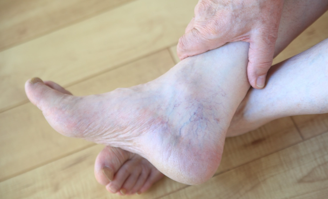
Sclerotherapy is a short procedure in which a fine needle is inserted into the target veins and a salt solution is injected. The chemical injection, which contains sclerosant, is administered into the vein, damaging and scarring it. The damaged vein then closes, causing it to fade away.

What can Sclerotherapy treat ?

Varicose veins often appear on older individuals of both genders. This procedure helps to reinvigorate confidence and self-esteem by eliminating unsightly veins that can cause embarrassment, particularly when wearing shorts, skirts, bathing suits and dresses. Sclerotherapy also brings a more youthful appearance back to the legs and feet.

While most patients will get sclerotherapy for cosmetic purposes – to eliminate the appearance of varicose and spider veins. The procedure also can improve related symptoms such as aching, swelling, burning and night cramps.

How are spider veins diagnosed?

Your dermatologist will perform a physical exam to diagnose spider veins, carefully examining the patterns of veins on your legs, feet, or other areas. The doctor will also check for any signs of swelling or changes in skin color. The dermatologist may also perform an ultrasound or venogram to see how blood is flowing through your veins.

What are my spider vein treatment options?

Based on the information from the physical exam and testing, your Forefront dermatologist will recommend the best treatment for spider veins. To provide you with the most effective results, your dermatologist may recommend more than one treatment option. Treatments range from self-care at home, spider vein treatment injection, and laser therapy.

Injection treatment: Sclerotherapy is a treatment that uses a series of injections to cause spider veins to collapse. The procedure doesn’t require anesthesia and is performed by a board-certified dermatologist at Forefront Dermatology.

Self-care: If you have small spider veins that aren’t causing your legs to ache or swell, following these self-care tips at home can improve circulation:

Exercise regularly:

  • Keep your legs elevated while sitting to prevent blood from pooling in your legs
  • Wear compression stockings to reduce swelling and improve blood flow
  • If you sit or stand for long periods, take a small break every 30 minutes to walk around
  • Avoid soaking in a hot bath for extended periods, which can cause your veins to swell

What happens during a sclerotherapy procedure?

During your sclerotherapy injection treatment session, your dermatologist will inject a solution directly into the affected veins. The chemical solution affects the walls of the vein, causing them to swell shut. Once the vein collapses, blood is prevented from flowing into the area. If you have several spider veins, the treatment may require multiple injections to target the wall of each vein. Depending on the number of veins requiring treatment, the procedure can last between fifteen minutes to one hour.

After injecting the affected veins, your dermatologist will massage the treatment area and place a compression stocking on each leg. To help with healing and to maintain pressure on the treated veins, your dermatologist will recommend wearing compression stockings for two to three weeks. It’s also important to take daily walks and move around as much as possible after the procedure to prevent blood clots from forming and ensure the best possible results. You can usually return to work and most everyday activities following your procedure. We also recommend avoiding direct sun exposure for at least two weeks.

Over three to six weeks, your spider veins will gradually fade. Most patients require two or three treatments for full result

How do Dermal Fillers work ?

Dermal fillers help to diminish facial lines and restore volume and fullness in the face. As we age, our faces naturally lose subcutaneous fat. The facial muscles are then working closer to the skin surface, so smile lines and crow’s feet become more apparent.

Dermal fillers differ in chemical make-up, longevity, and have varying degrees of softness. Softer fillers are used in the lips, for example, while sturdier fillers might be desired to enhance cheekbones.

Your surgeon will determine with you the best type and volume of filler needed for your particular areas of concern.

For many people, the use of “off-the-shelf” fillers can be a simple office-based procedure that can nicely enhance their appearance. These dermal fillers are very predictable, and have relatively minimal risks and side effects.

What can Dermal Fillers treat ?

Dermal fillers are commonly used as lip fillers to plump thin lips, enhance shallow contours, soften facial creases and wrinkles and improve the appearance of recessed scars.

What results can be expected from Dermal Fillers ?

In some situations, it may not be possible to achieve optimal results with a single procedure. Most fillers do dissipate over time. To maintain your correction, you will need to consider repeating the injection process at intervals.

There is no downtime with dermal fillers. You can resume most activities right away. Your initial appearance after treatment with any dermal filler may include:

  • An “over-filled” appearance to treated areas
  • Mild swelling or bruising
  • Temporary numbness or redness
  • These conditions can be alleviated with topical icing and will improve within a matter of hours or just a few days.
Young lady dermal fillers
Each year, over
1M Americans
get Botox injections
Botox is the
top nonsurgical
procedure in the U.S.
Botox is getting
more popular
each year in the U.S.
Botox Cosmetic is
FDA approved
for safety and efficiency

How Does BOTOX Work?

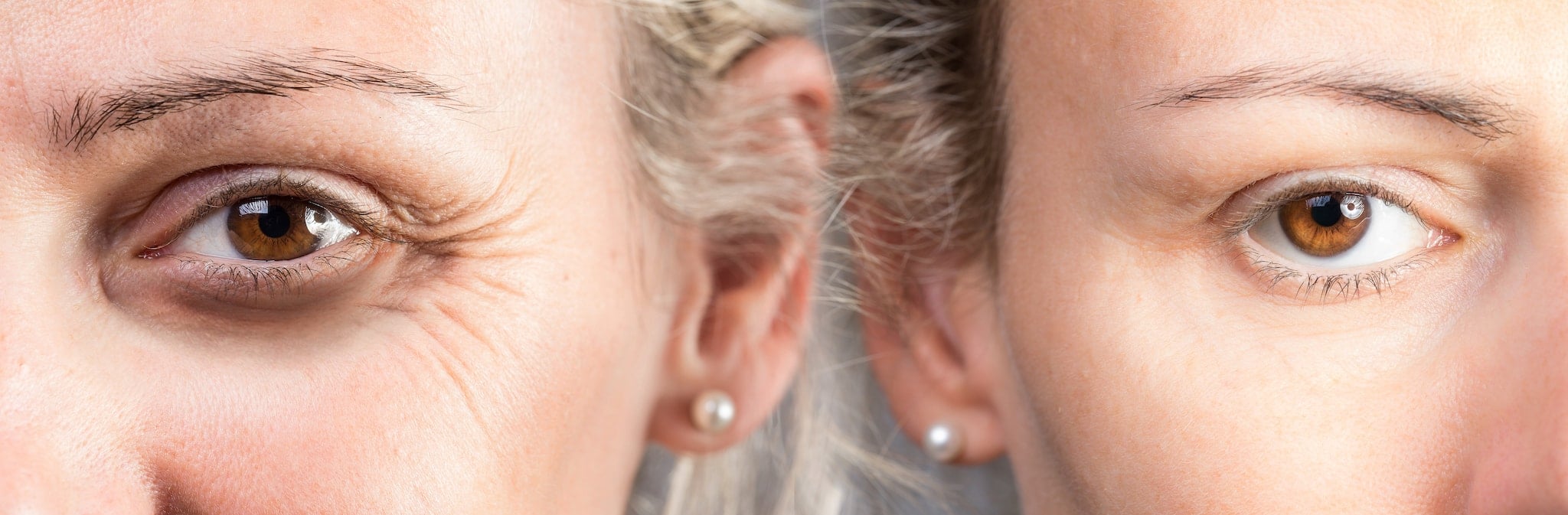
BOTOX Cosmetic is an injectable treatment that can soften the appearance of wrinkles, creating smoother and younger-looking skin. As you age, you might start to notice lines and wrinkles on your forehead, between your eyebrows, and around your eyes. Repetitive facial expressions, such as squinting, smiling, or frowning, can cause these wrinkles to deepen over time and remain noticeable even when your face is relaxed.

How Does BOTOX Cosmetic Treatment Work?

BOTOX contains purified botulinum A toxin, which temporarily blocks the nerve signals that cause facial muscles to contract. This reduction in muscle movement allows the skin to remain at rest even when making certain facial expressions, which smooths the lines and wrinkles for a more youthful look.

Before and After Comparison

Before and After

What Happens During a BOTOX Treatment Session?

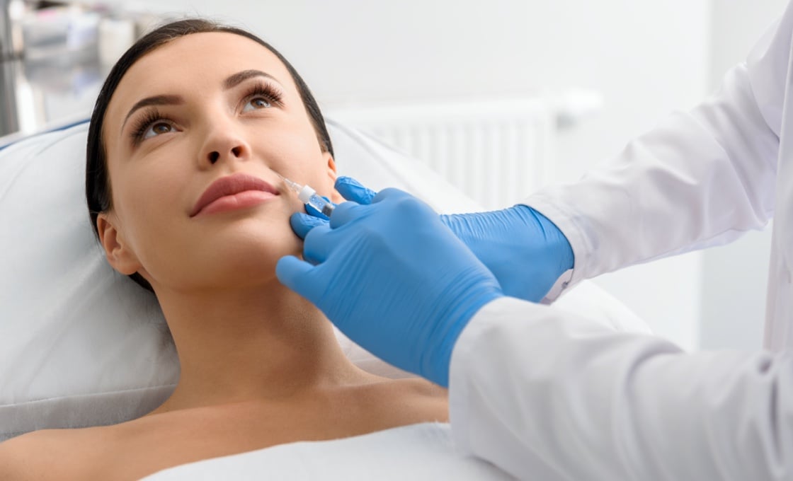
To ensure that you’re as comfortable as possible during the procedure, your Forefront dermatologist will apply ice or a topical anesthesia to numb the treatment areas. Next, they’ll prepare a small syringe with the recommended dosage of botulinum toxin. A thin needle will be used to inject the serum into the targeted areas. Most patients feel mild discomfort or a slight pinch during treatment.

It’s common for people to receive three to five injections per BOTOX treatment, but the number you receive will depend on the size of the treatment area and the severity of your symptoms. This non-invasive procedure is usually straightforward and relatively quick, and can be completed in as little as ten minutes.

Your expert Forefront dermatologist will give you instructions for care following your procedure, and you’ll be able to immediately resume your everyday activities. There is minimal to no downtime associated with the procedure, although you should be aware of certain safety restrictions. It’s important for you to avoid rubbing or massaging the treatment area since this can cause the botulinum toxin to migrate to other areas of the face.

What Are the Treatment Areas for BOTOX Injections?

Common treatment areas include wrinkles between the eyebrows, the corners of the eyes, the forehead, and the sides of the mouth. You can also use Botox to treat hyperhidrosis and migraines.

What Results Can I Expect from BOTOX?

Within 24 to 48 hours, you’ll notice a significant reduction of moderate to severe lines and wrinkles. Full results can be expected within one week. Your results will last for approximately four months as the ingredients are broken down and absorbed by the body.

To maintain your smooth, more youthful appearance, you’ll need to receive another treatment within three to six months. Since the formula is safe for repeated treatments, you can schedule injections with your Forefront dermatologist as needed. Our doctors will design a treatment plan that will best meet your aesthetic goals.

Are There Any Side Effects?

When performed by one of our Board-Certified Forefront dermatologists, any side effects are generally mild, and many people don’t experience any side effects at all. Some people might have redness or bruising at the injection site, although this is only temporary and usually subsides after a few days. Although rare, these serious side effects may occur:

  • Double vision
  • Blurred vision
  • Difficulty swallowing
  • Wheezing or difficulty breathing
  • Rash
  • Itching
  • Dizziness

You should seek emergency medical attention as soon as possible if you experience any side effects after receiving Botox.

Where Can I Receive Botox Treatment?

The Botox Cosmetic procedure is offered at many of our practice locations. Our Board – Certified dermatologists are experts in performing this procedure.